


Services on Demand
Article
 pdf in English
 Article in xml format
 Article references
 Send this article by e-mail
Related links
Share

 Permalink
RSBO (Online)
On-line version ISSN 1984-5685
RSBO (Online) vol.12 n.1 Joinville Jan./Mar. 2015
Original Research Article
Radiopacity analysis of humidity influence on the solubilization of different retrograde filling materials
Kiany Scarssi NunesI; Luiza Bonezi BoffI; Juliana RoyerI; Tiago André Fontoura de MeloI
I Dental School, College of Serra Gaúcha – Caxias do Sul – RS – Brazil
ABSTRACT
Introduction and Objective: This study aimed to analyze the level of radiopacity of different materials used in endodontic retrofilling procedure regarding the influence of humidity on solubilization over a period of 30 days. Material and methods: 10 specimens with 10 mm diameter and 1mm thick were made with each of the materials. The following retrograde filling materials were tested: glass ionomer Vitro Fil LC®, IRM®, MTA Angelus®, Sealer 26® and silver amalgam capsule DFL Alloy. These materials have been handled by a single operator, according to the recommendations of their respective manufacturers. The obtained specimens were surrounded by moist gauze and incubated in a heater for thirty days at 37°C and 100% humidity. The samples were x-rayed, via the digital system Digora Optime®, at three different moments: first, 15th, and 30th day of incubation. For the analysis of radiopacity of the samples, the shades of gray were measured through ImageTool® software. Results and Conclusion: Data were subjected to statistical analysis using ANOVA, repeated measures design, followed by Tukey test with a significance level of 5%. According to the results, it could be observed that the only tested materials that have undergone changes in radiopacity, in accordance with the period of incubation in humidity, were the glass ionomer Vitro Fil LC® and Sealer 26®.
Keywords: Endodontics; root canal filling materials; solubility.
Introduction
The retrograde filling is a procedure often used in paraendodontic surgery in order to seal the canal with at root resection in the apical portion of the tooth root19,22. To verify the correct filling of retrocavity and to facilitate surgery following-up, the retrofilling material must provide radiopacity enough to be distinguished from the surrounding anatomical structures18. According to Shah et al.16, the materials used in retrofilling must show a certain level of radiopacity that enables differentiating the root dentin from support bone.
Many materials have been tested and used in paraendodontic surgery, such as silver amalgam10, MTA5, glass ionomer13 and zinc oxide and eugenol-based cements7. But to date none behaved optimally.
The ideal retrofilling material should be easy to handle, have good radiopacity, provide dimensional stability in order to maintain a good apical sealing, be non-toxic to periapical tissues, and be insoluble or have low solubility11.
Thus, given that the used root-end filling materials are in close and continuous contact with the tissue fluids of the periapical region, this study aimed to analyze the influence of humidity on the level of radiopacity of some dental materials used in endodontic retrofilling.
Material and methods
The root-end filling materials tested were divided into five groups: group I – glass ionomer Vitro Fil LC® (DFL Produtos Odontológicos, Rio de Janeiro, Brazil), group II – IRM® (Dentsply/Maillefer Instruments S.A., Bal laigues, Switzerland), group III – MTA Angelus® (Angelus Indústria de Produtos Odontológicos Ltda., Londrina, Paraná, Brazil), group IV – Sealer 26® (Dentsply/Maillefer Instruments S.A., Ballaigues, Switzerland), and group V – silver amalgam capsules DFL Alloy (DFL Produtos Odontológicos, Rio de Janeiro, Brazil).
Ten specimens with 10 mm diameter and 1 mm thick were prepared with aid of a flexible silicone condensation matrix (Vigodent S.A. Indústria e Comércio, Bonsucesso, Rio de Janeiro, Brazil), for each of the materials. The root-end filling materials were handled by a single operator and according to the recommendations of the respective manufacturers.
The samples during the experiment, were wrapped in moist gauze and incubated for 30 days, at 37ºC and 100% humidity, in a bacteriological incubator403/N (Multitec Equipamentos para Laboratór io, Canoas, Rio Grande do Sul, Brazil).
To carry out the radiographic images, the samples were placed on an image plate (sensor) digital system Digora Optime® (Soredex Corp., Tuusula, Finland). This sensitized plate, after the radiographic shot with x-ray machine X Gnatus (70kVp – 7mA, Gnatus Equipamentos Odontológicos Ltda., Ribeirão Preto, São Paulo, Brazil), was introduced in laser optical reader of Digora Optime® system in order to obtain the desired image with an exposure time of 0,32 seconds at a distance of 30 cm. For standardization of distance and position of images a customized to a radiographic platform previously was made.
The radiographic images of the specimens were made at three different times: 1st, 15th, and 30th day after incubation in the heater. In total, 150 radiographic images were made.
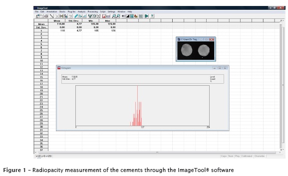
To analyze the radiopacity the grayscale of the images were measured through the ImageTool® software (UTHSCSA, San Antonio, Texas). To measure the shades of gray, ranging from 0 to 255 pixels, the “histogram” was used in a standard area of 20 x 19 pixels positioned in a standardized manner at the central area of the images (figure 1).
The data obtained in the assessment were treated and analyzed by ANOVA statistical analysis, using the repeated measure design, following by Tukey test for multiple comparisons, at a significance level of 5%.
Results
The results with the mean and standard deviation of radiopacity in pixels of the five root-end filling materials are shown in table I.
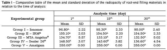
Means followed by different capital letters in line differ significantly by ANOVA using repeated measure design, followed by the Multiple Comparison Tukey Test, at a significance level of 5%.
Discussion
The search for the ideal biomaterial for paraendodontic surgery is a constant in modern Endodontics, since it is difficult to obtain in a single product all the desired physical-chemical, mechanical and biological.
The difference in radiopacity between the different retrofilling materials employed in the paraendodontic surgery exposes some deficiency in the existing products on dental market, which can serve as exclusion criteria in clinical choice for the material to be used with this purpose.
Thus, the radiopacity has received attention in several studies4,18,24. Some studies1,2 have used photodensitometry and aluminum scales to compare the radiopacity of sealers. However, in recent years, evaluations from digital radiographic images comparing or not to the aluminum scales have been quite frequent ly3,23. With the advent of digital images, this type of assessment has become more effective and fast, since the radiographic density is directly obtained, because the pixels already have their certain shades of gray.
Based on the assessment of the gray tones, it could be appreciated in this study statistical difference in the radiopacity only for following the root-end filling materials: glass ionomer Vitro Fil LC® and Sealer 26®.
The solubilization and disintegration of the materials allow the appearance of voids within the obturator mass, promoting retrograde infiltration of tissue fluids, which may compromise the sealing and the treatment success.
In the studies of Fidel et al.9, Tanomaru Filho et al.20, Scelza et al.15, and Kuga et al.14, Sealer 26® showed low solubility index, corroborating the results obtained in this present study. Sealer 26® is an epoxy resin-based cement containing calcium hydroxide in its composition. By having its sealing ability compared with the use of IRM® and glass ionomer cement, with the specimens filled with these materials in contact with human saliva for 60 days, it was observed that Sealer 26® showed excellent ability to seal when used as retrograde filling material, as well as great capacity to prevent bacterial leakage 17.
With regard to glass ionomer, Carvalho Júnior et al.6 studied the solubility, disintegration and dimensional changes of Ketac-ENDO® compared to Endofill® and Sealer 26®. They verified that Endofill® and Ketac-ENDO® had higher solubility values and disintegration than those recommended by the ADA specification. According to Gorodovsky and Zidan12, the glass ionomer presented as inherent characteristics: solubility and power of disintegration in liquids and wet media.
Concerning to the silver amalgam capsules DFL Alloy, IRM®, and MTA Angelus® did not show radiopacity alterations at the analyzed periods. Although the silver amalgam has presented satisfactory results for radiopacity, it has some limitations, such co mo high power of oxidation and tissue contamination by mercury11. According to the study of Crooks et al.8, zinc oxideeugenol- based materials, such as IRM®, have good mechanical resistance and low solubility. Notwithstanding, according to Torabinejad et al.21, MTA, in addition to biocompatibility, favors the formation of hard tissue after paraendodontic surgery, and does not exhibit, after setting, solubility in the presence of humidity, and thus is the best material for use in retrograde fillings.
Conclusion
According to the results found, it could be seen that the glass ionomer Vitro Fil LC® and Sealer 26® underwent radiopacity alterations, according to the period of humidity incubation. Concerning to IRM®, MTA Angelus® and silver amalgam DFL Alloy did not undergo modifications.
References
1. Almeida PM, Antonio MPS, Moura AAM. Estudo comparativo da radiopacidade de quatro cimentos obturadores de canais radiculares. Rev Inst Ciênc Saúde. 1998;16(1):27-30. [ Links ]
2. Beyer-Olsen EM, Orstavik D. Radiopacity of root canal sealers. Oral Surg Oral Med Oral Pathol. 1981;51(3):320-8.
3. Bicheri SAV, Victorino FR. Comparative evaluation of radiopacity of MTA Fillapex® endodontic sealer through a digital radiograph system. RSBO. 2013;10(2):149-52.
4. Bortoluzzi EA, Guerreiro-Tanomaru JM, Tanomaru-Filho M, Duarte MA. Radiographic effect of different radiopacifiers on a potential retrograde filling material. Oral Surg Oral Med Oral Pathol Oral Radiol Endod. 2009;108(4):628-32.
5. Cami l leri J, Sorrent ino F, Damidot D. Investigation of the hydration and bioactivity of radiopacified tricalcium silicate cement, Biodentine and MTA Angelus. Dent Mater. 2013;29(5):580- 93.
6. Carvalho Júnior JR, Guimarães LF, Correr- Sobrinho L, Pécora JD, Sousa-Neto MD. Evaluation of solubility, disintegration, and dimensional alterations of a glass ionomer root canal sealer. Braz Dent J. 2003;14(2):114-8.
7. Chong BS, Pitt Ford TR, Hudson MB. A prospective clinical study of mineral trioxide aggregate and IRM when used as root-end filling materials in endodontic surgery. Int Endod J. 2003;36(8):520-6.
8. Crooks WG, Anderson RW, Powell BJ, Kimbrough WF. Longitudinal evaluation of the seal of IRM root end fillings. J Endod. 1994;20(5):250-2.
9. Fidel RAS, Spanó JCE, Barbin EL, Silva RG, Pécora JD. Estudo in vitro sobre a solubilidade e a desintegração de alguns cimentos endodônticos que contêm hidróxido de cálcio. Rev Odontol Univ São Paulo. 1994;8(3):217-20.
10. Frank AL, Glick DH, Patterson SS, Weine FS. Long-term evaluation of surgically placed amalgam fillings. J Endod. 1992;18(8):391-8.
11. Gartner AH, Dorn SO. Advances in endodontic surgery. Dent Clin North Am. 1992;36(2):357- 78.
12. Gorodovsky S, Zidan O. Retentive strength, disintegration, and marginal quality of luting cements. J Prosthet Dent. 1992;68(2):269-74.
13. Jesslén P, Zetterqvist L, Heimdahl A. Longterm results of amalgam versus glass ionomer cement as apical sealant after apicectomy. Oral Surg Oral Med Oral Pathol Oral Radiol Endod. 1995;79(1):101-3.
14. Kuga MC, Campos EA, Sant’Anna Júnior A, Vasconcelos FL, Silva AN, Nascimento CA. Avaliação do pH, da solubilidade e da infiltração marginal em retrobturações com o Sealer 26® puro ou acrescido de iodofórmio. RSBO. 2010;7(4):389-95.
15. Scelza MFZ, Scelza P, Costa RF, Câmara A. Estudo comparativo das propriedades de escoamento, solubilização e desintegração de alguns cimentos endodônticos. Pesq Bras Odontoped Clín Integr. 2006;6(3):243-7.
16. Shah PM, Chong BS, Sidhu SK, Ford TR. Radiopacity of potential root-end filling materials. Oral Surg Oral Med Oral Pathol Oral Radiol Endod. 1996;81(4):476-9.
17. Siqueira Júnior JF, Rôças IN, Abad EC, Castro AJ, Gahyva SM, Favieri A. Ability of three root-end filling materials to prevent bacterial leakage. J Endod. 2001;27(11):673-5.
18. Tagger M, Katz A. A standard for radiopacity of root-end (retrograde) filling materials is urgently needed. Int Endod J. 2004;37(4):260-4.
19. Tanomaru Filho M, Luis MR, Leonardo MR, Tanomaru JM, Silva LA. Evaluation of periapical repair following retrograde filling with different root-end filling materials in dog teeth with periapical lesions. Oral Surg Oral Med Oral Pathol Oral Radiol Endod. 2006;102(1):127-32.
20. Tanomaru Filho M, Moraes IG, Duarte MAH, Arekaki OT, Nishiyama CK. Avaliação do selamento apical de dois cimentos endodônticos à base de hidróxido de cálcio. Rev Bras Odont. 1996;53(3): 2-4.
21. Torabinejad M, Hong CU, McDonald F, Pitt Ford TR. Physical and chemical properties of a new root-end filling material. J Endod. 1995;21(7): 349-53.
22. Torabinejad M, Pitt Ford TR, McKendry DJ, Abedi HR, Miller DA, Kariyawasam SP. Histologic assessment of mineral trioxide aggregate as a root-end filling in monkeys. J Endod. 1997;23(4): 225-8.
23. Vidotto APM, Cunha RS, Zeferino EG, Rocha DGP, Martin AS, Bueno CES. Comparison of MTA Fillapex radiopacity with five root canal sealers. RSBO. 2011;8(4):404-9.
24. Vivan RR, Ordinola-Zapata R, Bramante CM, Bernardineli N, Garcia RB, Hungaro Duarte MA et al. Evaluation of the radiopacity of some commercial and experimental root-end filling materials. Oral Surg Oral Med Oral Pathol Oral Radiol Endod. 2009;108(6):35-8.
 Corresponding author:
Tiago André Fontoura de Melo
Rua Nicola Mathias Falci, n. 151 / casa 16 – Jardim do Salso
CEP 91410-330 – Porto Alegre – RS – Brazil
E-mail: tafmelo@gmail.com
Received for publication: November 19, 2014
Accepted for publication: December 22, 2014
